Bi2S3-SiO2 NRs/二氧化硅涂層硫化鉍納米棒多模態顯影劑可用于胃腸道GI成像技術
利用硫酸鋇懸浮液作為X光顯影劑用于小腸成像,由于其自身具備的非降解及其他一些不理想的特點使其在胃腸道穿孔及腸道結構觀測方面受到很大限制;其他常用的胃腸道顯影劑便是碘代分子,但因其本身X射線吸收率較低,所以通常需要大劑量使用才能達到理想的效果,這常常會使病人產生碘過敏性反應。
制備得到二氧化硅涂覆的硫化鉍納米棒(Bi2S3@SiO2 NRs)作為多模態顯影劑用于胃腸道的無侵害性實時成像以及直接觀測其在胃腸道下部的流經過程(Scheme 1)。經二氧化硅包覆后,在胃及小腸中,Bi2S3@SiO2 NRs展現出非常好的水溶性,生物相容性以及穩定性。通過TEM可以看出,Bi2S3 NRs寬約10 nm,長約50 nm,元素分析譜圖顯示成功制備得到高純度的Bi2S3 NRs(Fig. 1),經二氧化硅涂覆后,Bi2S3@SiO2 NRs呈現單分散性,SiO2殼層厚度約為6 nm(Fig. 2)。
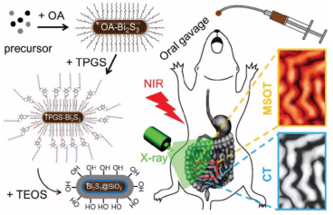
Scheme 1 Schematic illustration of the preparation of Bi2S3@SiO2 NRs for bimodal CT/PAT imaging of the GI tract principle based on the unique properties of Bi2S3@SiO2 NRs。
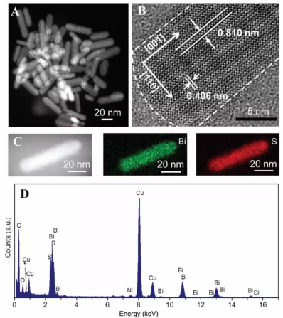
Fig. 1 Characterization of Bi2S3 NRs. (A) HAADF-STEM image and (B) HRTEM image of Bi2S3 NRs prepared by the solvothermal method. (C) Corresponding element mapping for Bi and S of the as-prepared Bi2S3 NRs. (D) EDS of the as-prepared Bi2S3 NRs.
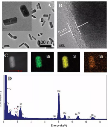
Fig. 2 Characterization of Bi2S3@SiO2 NRs. (A) TEM image and (B) HRTEM image of as-prepared Bi2S3@SiO2 NRs. Inset: HAADF-STEM image of Bi2S3@SiO2 NRs. (C) Corresponding element mapping for Bi, S and Si of Bi2S3@SiO2 NRs. (D) EDS of Bi2S3@SiO2 NRs.
通過CT成像分析發現,隨著Bi2S3@SiO2 NRs濃度增加,其HU值明顯增加,相同濃度下的HU值明顯高于硫酸鋇,PAT信號隨濃度增加也呈線性增長關系(Fig. 3)。
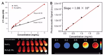
Fig. 3 CT and PAT phantom images of Bi2S3@SiO2 NRs with different concentrations in vitro. (A) Plot of Hounsfield units (HU) values and of Bi2S3@SiO2 NRs and BaSO4 suspension versus the sample concentrations and CT phantom images of Bi2S3@SiO2 NRs and BaSO4 suspension samples with different concentrations. (B) Plot of the photoacoustic signal versus Bi2S3@SiO2 NRs concentrations and PAT phantom images of Bi2S3@SiO2 NRs aqueous solutions with different concentrations.
對Bi2S3@SiO2 NRs的生物相容性進行檢測,發現16HBE與Bi2S3@SiO2 NRs共培養24 h后并未顯示出明顯的毒性,并且該粒子進入秀麗隱桿線蟲體內后對其壽命也沒有明顯的影響,這就說明Bi2S3@SiO2 NRs具有非常好的生物相容性(Fig. 4)。
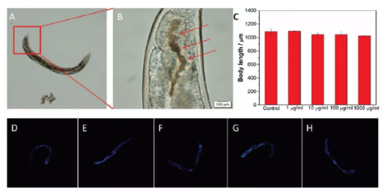
Fig. 4 Biosafety assessment of Bi2S3@SiO2 NRs by the C. Elegans model. (A) Bright field image of the NRs distribution in the GI tract of C. Elegans. Worms feed on NGM plates with Bi2S3@SiO2 NRs (1000 μg mL?1) transferred onto an agar pad after 1 h. (B) The distribution of food containing Bi2S3@SiO2 NRs (red arrows) in the intestine of the worm’s tail. (C) Effects of Bi2S3@SiO2 NRs with different concentrations on body length of C. Elegans. (D–H) Effects of Bi2S3@SiO2 NRs treatments on the accumulation of lipofuscin in age-synchronized worms. Representative fluorescent images of worms fed with 0, 1, 10, 100 and 1000 μg mL?1 Bi2S3@SiO2 NRs, respectively.
Bi2S3@SiO2 NRs以口服的方式進入BALB/c裸鼠體內,通過CT及PAT對Bi2S3@SiO2 NRs進行實時成像追蹤,發現該粒子通過胃腸道漸漸進入小腸較后以糞便的形式排出體外,該過程中對胃腸道的功能沒有明顯的損傷,說明Bi2S3@SiO2 NRs對組織無侵入性損傷(Fig. 5, 6, 7)。
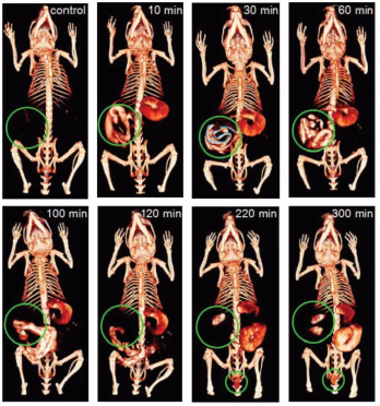
Fig. 5 CT imaging of the GI tract in vivo. In vivo X-ray CT imaging of the GI tractin BALB/c nude mice at different intervals after oral administration of Bi2S3@SiO2 NRs.
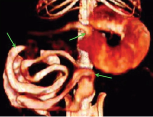
Fig. 6 Enlarged images of CT images of the GI tract of mice 30 min post oral administration of Bi2S3@SiO2 NRs.
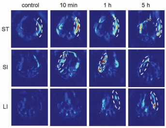
Fig. 7 PAT imaging of the GI tract in vivo. PAT cross-sectional image of the GI tract of BALB/c nude mice at different intervals after oral administration of Bi2S3@SiO2 NRs: stomach (ST), small intestine (SI) and large intestine
wyf 04.02


 齊岳微信公眾號
 官方微信
官方微信 庫存查詢
庫存查詢